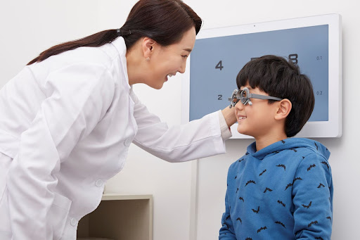
Our paediatric ophthalmologists and surgeons at ParkCity Medical Centre (PMC) can help diagnose and treat childhood eye disorders to ensure that your child has the best possible vision.
During the first year of life, your child's eyes should be examined by a paediatrician. If you or your paediatrician see anything out of the ordinary or if you have a family history of eye disorders, you must bring your child to an ophthalmologist.
All children, even those with good vision, are advised to undergo a comprehensive eye exam by their fourth birthday and every two years thereafter. Accurate diagnosis and appropriate treatment are the most effective ways to safeguard or restore your child's vision.
The above is recommended as some babies and children suffer from retinal conditions due to genetic disorders or eye injuries. At PMC, we address a variety of retinal issues in children. These include:
-
Retinal Detachment
Retinal detachments happen when the retina pulls away from its normal position. This can occur in children for a variety of causes, including:
-
Complications following eye surgery, especially cataract surgery
-
Family history of detached retinas
-
Prior detachment in one eye
-
Prior eye disease or inflammation
-
Severe near-sightedness
-
Traumatic eye injury
Symptoms may include:
-
Blurred vision
-
Curtain-like shadow over the visual field
-
Increasingly worse side vision
-
Sudden flashes of light or floaters – dark spots drifting across the line of vision
The longer a retinal detachment remains untreated, the more likely for permanent visual loss to occur.
-
Retinopathy of Prematurity (ROP)
ROP is a condition that affects babies who are born prematurely or with low birth weight. Premature newborns are typically born before their retinal blood supply has fully developed.
As their eyes develop new blood vessels, these veins bleed and grow into areas of the eye where they are not supposed to be. This results in scar tissue within the eyes. Without treatment, these infants are at risk of developing retinal detachment, visual loss and eventually, blindness.
-
Retinoblastoma
Retinoblastoma is a fast-growing tumour that develops from immature retinal cells, the light-detecting tissue of the eye. It is the most common ocular tumour in children, occurring in approximately 1 in 14,000–18,000 live births. As a result, an estimated 8000 cases will occur globally. Adults, on the other hand, are extremely unlikely to have this form of cancer.
Common Eye Tests
Before a diagnosis can be made, our ophthalmologists will conduct a series of tests to have a better understanding of the condition and how far it has progressed. Tests include:
-
Retinal examination – As part of a comprehensive eye exam, your child's doctor may use a bright light and a special lens (ophthalmoscope) to examine the retina. This provides the doctor with a highly thorough picture of any retinal abnormalities.
-
Ultrasound imaging – If there is bleeding inside the eye, the doctor may utilise ultrasound imaging to detect any abnormalities in the retina.
Treatments
Once diagnosed, our ophthalmologists will recommend a treatment that fits your child the best. Among the treatments are:
-
Pneumatic retinopexy – In this procedure, the surgeon injects gas or air bubble into the centre of the eye to press the retinal hole against the eye's wall, preventing intraocular fluid from flowing behind the retina. Any retinal hole or tear is also sealed with laser coagulation (heat) or cryopexy (freezing). Both procedures prevent fluid from seeping beneath the retina. Any leftover fluid under the retina and the bubble Is reabsorbed over time, and the retina reattaches to the eyewall.
-
Scleral buckle – A thin silicone band is sewn to the white (sclera) part of the eye during this treatment. The buckle will create an indentation in the eyewall, lessening the pressure created by the vitreous (a gel-like substance inside the eye) and preventing it from dragging on the retina. If there are multiple retinal holes, tears or a serious detachment, the surgeon may use a scleral buckle to wrap the entire eye. The buckle is invisible and is positioned such that it does not hinder vision.
-
Vitrectomy – A vitrectomy removes the vitreous fluid as well as the tissue that is dragging on the retina. The retina is then held in place by inserting air, gas or silicone oil into the vitreous area. It is reabsorbed over time and the vitreous gap will be refilled with natural fluid. A vitrectomy is sometimes paired with a scleral buckle in some patients.
At PMC, our highly trained experts include some of Malaysia's best ophthalmologists, who provide cutting-edge care through first-rate clinical practice and innovative medical diagnostics. At our new Eyecentric, our team of highly skilled eye surgeons also provides a variety of procedures, ranging from cataract surgery to the treatment of retinal problems such as retinal detachment and macular degeneration.
Meet our Specialist
Dr Ronald Arun Das
Designation
Consultant Ophthalmologist and Vitreo Retinal Surgeon
Dr V. Ulagantheran Viswanathan
Designation
Consultant Ophthalmologist and Vitreo Retinal Surgeon


